
Call us now :08071792071
 Send Inquiry
 Send Inquiry
MAGNUS TRINOCULAR INVERTED FLUORESCENCE MICROSCOPE FOR BRIGHT FIELD , PHASE CONTRAST & FLUORESCENCE APPLICATION MODEL FL INVERSE
Price 1126990.0 INR/ Piece
MOQ : 1 Piece
MAGNUS TRINOCULAR INVERTED FLUORESCENCE MICROSCOPE FOR BRIGHT FIELD , PHASE CONTRAST & FLUORESCENCE APPLICATION MODEL FL INVERSE Specification
- Focus System
- Coaxial coarse & fine focusing mechanism with tension adjustment
- Features
- Multi-mode application (bright field, phase contrast, fluorescence), modular illumination, ergonomic design, high stability
- View Head
- Trinocular, Siedentopf, 360 rotatable with diopter adjustment
- Spare Parts
- Extra bulb, fuses, objectives, eyepieces, stage clips, filters
- Theory
- Inverted fluorescence microscope suitable for bright field, phase contrast, and fluorescence applications.
- Drawtube
- Trinocular, 30 inclined
- Sensor
- Not applicable (optical system)
- Resolution
- High resolution, suitable for microbiology and cell culture observation
- Interface
- Trinocular phototube for camera or imaging device attachment
- Frame Rate
- Not applicable (optical system)
- Focal Distance
- 160mm (optical tube length)
- Magnification
- 40X to 400X (objective and eyepiece dependent)
- Dimensions
- Approx. 570mm (H) x 285mm (W) x 540mm (D)
- Focus Range
- Coarse and fine focusing with graduated adjustment
- Eyepieces
- WF10X (Wide Field, 22mm field of view)
- Eyepiece Tube
- Trinocular (two viewing, one photo port)
- Illumination
- 6V/30W Halogen Lamp (Transmitted), 100W Mercury Fluorescence Lamp (Incident)
- Coarse Adjustment Range
- 15mm or more
- Fine Adjustment Range
- 0.002mm precision
- Working Stage
- Mechanical XY stage with specimen holder, stage size approx. 242 x 208 mm
- Still Image Capture Resolution
- Camera dependent (compatible with high-resolution cameras)
- Video Capture Resolution
- Camera dependent (compatible with high-definition cameras)
- Image Format
- Compatible with standard microscopy camera adapters
- Interpupillary Distance
- 50-75 mm adjustable
- Objective Achromatic
- Plan Achromatic Objectives: 4X, 10X, 20X (Phase), 40X (Phase)
- Condenser
- Long Working Distance (LWD) Condenser for phase contrast, NA 0.3
- Light Source
- Transmitted: Halogen 6V/30W; Fluorescence: 100W Mercury lamp
- Specimen Holder
- Adjustable universal holder to accommodate Petri dishes, multi-well plates, flasks, or slides
- Color Correction
- Plan Achromatic objectives with improved color correction for fluorescence
- User Manual
- Comprehensive multilingual manual included
- Available Filters
- B/G/U/V multiband fluorescence filters supplied
- Stage Movement
- Precision mechanical, smooth XY movement with calibration scale
- Anti-Fungal Coating
- Optics and mechanical parts treated for anti-fungal protection
- Operating Environment
- Temperature 10-40C, relative humidity 30-85%
- Lamp Housing
- Integrated heat-resistant lamp housing for mercury lamp
- Safety
- Shielded lamp housing and UV protection features
- Lamp Life
- Halogen >2000 hours; Mercury >300 hours
- Power Supply
- AC 220V/110V, 50-60Hz compatible
- Filter Holder
- Equipped with slider type filter holder for quick exchange of barrier and excitation filters
MAGNUS TRINOCULAR INVERTED FLUORESCENCE MICROSCOPE FOR BRIGHT FIELD , PHASE CONTRAST & FLUORESCENCE APPLICATION MODEL FL INVERSE Trade Information
- Minimum Order Quantity
- 1 Piece
- Delivery Time
- 5 Week
- Main Domestic Market
- All India
About MAGNUS TRINOCULAR INVERTED FLUORESCENCE MICROSCOPE FOR BRIGHT FIELD , PHASE CONTRAST & FLUORESCENCE APPLICATION MODEL FL INVERSE
MAGNUS TRINOCULAR INVERTED FLUORESCENCE MICROSCOPE FOR BRIGHT FIELD
PHASE CONTRAST & FLUORESCENCE APPLICATION MODEL FL INVERSE
Infinity Corrected Optical System with inclined Trinocular Observation tube with 50:50 light path
having Interpupillary distance adjustment range of 5075mm, Plan Achromat objective of 4x, LWD
Plan Achromat Phase Objective of 10x (for BF & Phase), LWD Semi Plan Apo Phase 20x (for BF/
PH/ FL), LWD Semi Plan Apo Phase 40x (for BF/ PH/FL), quintuple revolving nosepiece, LWD
Condenser with NA 0.30, WD 72 mm, Transmitted LED illumination, 215 X 250 mm fixed stage with
Attachable mechanical stage having movement of 120 X 80 mm. LED based three band switching
Fluorescence Attachment for Blue Filter (LED Module 470 nm) Green Filter (LED Module 560 nm ) &
UV Filter (LED Module 385 nm) for FL Inverse.
Versatile Imaging Modes
This microscope supports bright field, phase contrast, and fluorescence observation. Users can efficiently switch between these techniques to study cell cultures and microorganisms, gaining comprehensive insights. Its modular illumination system, with both halogen and mercury lamps, enhances versatility for diverse laboratory protocols.
Precision Engineering and Ergonomics
Equipped with a precision mechanical stage, smooth XY movement, and calibrated scales, the Model FL Inverse provides high stability and accuracy. The coaxial coarse and fine focusing mechanism ensures precise adjustments, while the ergonomic, trinocular Siedentopf head promotes user comfort during long observation periods.
Enhanced Safety and Durability
Safety is prioritized with shielded, heat-resistant lamp housing and UV protection features. Optics and mechanical components are treated with an anti-fungal coating, making the system durable and long-lasting, even in humid laboratory environments. The microscope adheres to stringent safety and operational standards.
FAQ's of MAGNUS TRINOCULAR INVERTED FLUORESCENCE MICROSCOPE FOR BRIGHT FIELD , PHASE CONTRAST & FLUORESCENCE APPLICATION MODEL FL INVERSE:
Q: How do I exchange the fluorescence filters on the MAGNUS FL Inverse microscope?
A: The microscope is equipped with a slider-type filter holder, enabling quick and simple exchange of multiband B/G/U/V fluorescence filters. Simply slide the filter holder to select the desired barrier or excitation filter for your specific fluorescence application.Q: What specimens can be accommodated on the stage of this microscope?
A: The microscope includes an adjustable universal specimen holder that can securely accommodate various laboratory vessels such as Petri dishes, multi-well plates, flasks, and standard microscope slides, providing flexibility for a broad range of sample types.Q: When should the microscope's lamps be replaced, and how long do they last?
A: The halogen lamp offers a lifespan of over 2,000 hours, while the mercury fluorescence lamp provides over 300 hours. It's recommended to replace lamps once they reach their rated lifespan to maintain optimal performance. Extra bulbs are supplied as spare parts.Q: Where can I attach a camera or imaging device for documentation?
A: A trinocular phototube is provided, allowing users to attach a compatible camera or imaging device. This enables high-resolution still image and video capture for thorough documentation and analysis of microscopy results.Q: What is the process for achieving optimal phase contrast and fluorescence imaging?
A: To achieve optimal imaging, adjust the long working distance condenser for phase contrast, select the correct objective, and ensure the specimen is properly positioned. Use the fine focus for precision adjustment, and switch filters as needed for fluorescence detection of your target markers.Q: How does the anti-fungal treatment benefit the microscope's longevity?
A: Anti-fungal coatings applied to the optics and mechanical parts prevent fungal growth, particularly in areas with high humidity. This protection significantly extends the service life and maintains the optical performance of the microscope over time.Q: What are the main advantages of using this model for cell culture observation?
A: This model provides high-resolution imaging, ergonomic operation, and versatile specimen handling, making it ideal for cell culture and microbiology studies. Enhanced color correction, phase contrast, and fluorescence options offer detailed observation and analysis of live cells and cultures.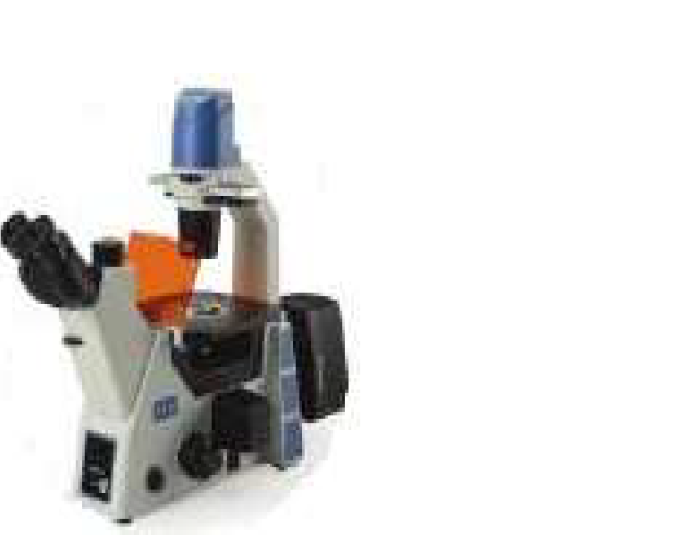
Tell us about your requirement

Price:
Quantity
Select Unit
- 50
- 100
- 200
- 250
- 500
- 1000+
Additional detail
Mobile number
Email

More Products in Inverted Microscope Category
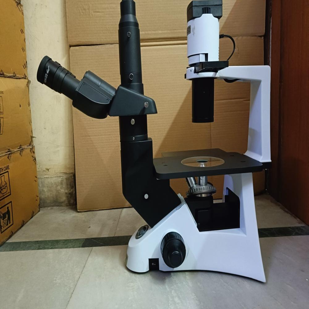
MAGNUS INVERTED TISSUE CULTURE TRINOCULAR MICROSCOPE MODEL INVI (LED)
Price 209970.0 INR / Set
Minimum Order Quantity : 1 Set
Features : LED illumination, modular stage design, ergonomic operation, suitable for Petri dishes/flasks, antifungal optics
Interpupillary Distance : 50 75mm adjustable
Eyepiece Tube : Trinocular, 45 inclined
Dimensions : Approx. 460mm x 380mm x 580mm
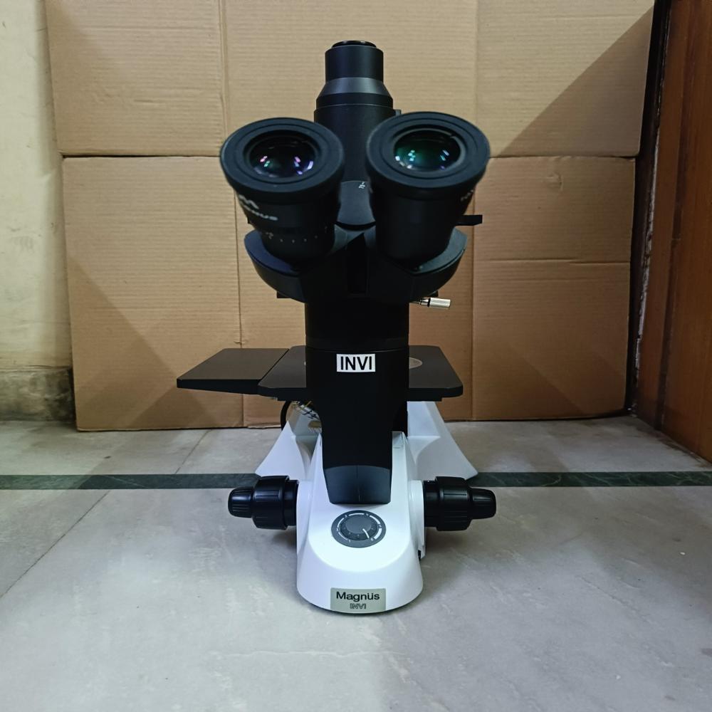
MAGNUS INVERTED TISSUE CULTURE TRINOCULAR MICROSCOPE MODEL INVI (HL)
Price 204330.0 INR / Piece
Minimum Order Quantity : 1 Piece
Features : Long working distance objectives, antifungus coated optics, suitable for Petri dishes and culture flasks, phase contrast optional.
Interpupillary Distance : Adjustable from 48mm to 75mm.
Eyepiece Tube : Trinocular; interpupillary adjustment 48mm75mm.
Dimensions : Approx. 490 mm (H) x 220 mm (W) x 510 mm (D).
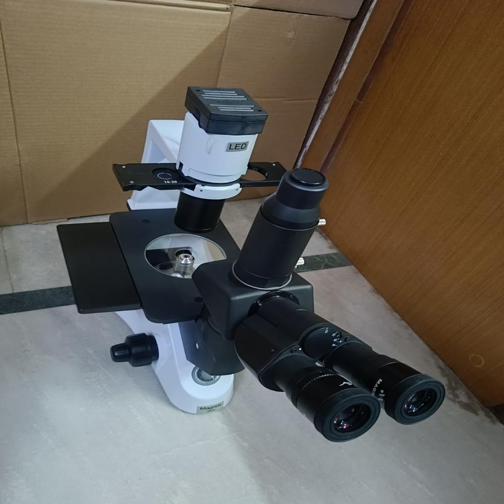
MAGNUS INVERTED TISSUE CULTURE TRINOCULAR MICROSCOPE MODEL INVI PLUS (HL)
Price 239000.0 INR / Piece
Minimum Order Quantity : 1 Piece
Features : Stable metal frame, ergonomic handle, antifungus treated optics, long working distance for culture flasks and Petri dishes.
Interpupillary Distance : Adjustable 55 mm to 75 mm.
Eyepiece Tube : Inclined 45, rotatable 360 trinocular tube with interpupillary distance adjustment.
Dimensions : Approx. 490 x 260 x 530 mm.
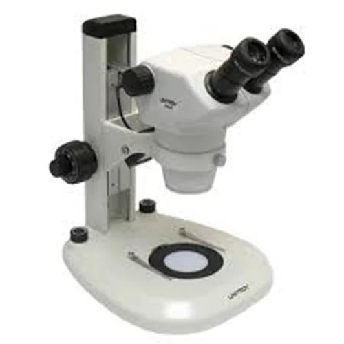
MAGNUS INVERTED TISSUE CULTURE TRINOCULAR MICROSCOPE MODEL INVI (LED) WITH ACCESSORIES:
Price 283755.0 INR / Set
Minimum Order Quantity : 1 Piece
Features : High quality


 Send Inquiry
Our Products
- Digital Colorimeter
- Visible Spectrophotometer
- Trinocular Microscope
- Biological Microscope
- Microscope Cmos Camera
- Physics Equipments
- Binocular Microscope
- Lab Equipments
- Monocular Microscope
- Research Microscopes
- Lab Reagents
- Inverted Microscope
- Turbidity Meter
- Stereoscopic Microscope
- Multi Head Teaching Microscope With LED
- Digital Melting Point Apparatus
- Stainless Steel Vertical Autoclave
- Laboratory Chemicals
- Auto Universal Titrator
- UV VIS Spectrophotometers
- Analytical And Scientific Instruments
- Digital Ph Meter
- Conductivity Meter
- Water And Soil Testing Kit
- Digital TDS Meter


Flat No. G-3, Plot No. 286, Sector-4, Vaishali,Ghaziabad - 201019, Uttar Pradesh, India

Mr. Pankaj Saran
(Proprietor )
Mobile :08071792071
 Send Inquiry
 Send SMS
 Call Me Free
Developed and Managed by Infocom Network Private Limited.
